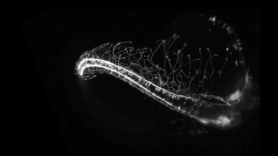
Nervous system captured growing in incredible timelapse footage (VIDEO)

Unbelievable 3D footage, capturing the meticulous development of a zebrafish embryo’s nervous system, has won a major photography award. The mesmerising microscope video was filmed over a 16-hour period.
The fascinating timelapse video, shot by Dr. Elizabeth Haynes and Jiaye ‘Henry’ He, won the 2018 Nikon Small World in Motion competition. Haynes studies the role of kinesin light chain genes during the highly complex development of sensory neurons, while He develops microscopy technology to capture the incredible process.
Together, the pair filmed the mesmerizing process inside their home-built microscope. The embryo grew in water - a phenomenon which is incredibly challenging to capture on camera as the specimen can easily move out of shot. However, the alternative technique to mount the zebrafish to a block of gel to restrict its movement could result in a less accurate portrayal of its neuron development.
"There was an amount of luck present for it to remain in a good position during the entire movie," Haynes explained to Live Science.
“I hope people see this video and understand how much we share with other organisms in terms of our development,” said Haynes of the striking, almost sci-fi-like footage.
“A neuron is a neuron, and it’s really amazing how, most of the time, development goes right when so much could go wrong. There is so much art occurring within science and nature, and it’s really special to watch.”
READ MORE: ‘Invasion of the body snatchers’: Mind-control fungus shows flies a gruesome end
Haynes says there is still much be to learned about the roles of kinesin light chain genes, and a better understanding of the changes in their growth could help neurodegenerative disorders such as Alzheimer's disease.
Think your friends would be interested? Share this story!


